Introduction to Cervical Decompression
Cervical decompression surgery relieves severe pressure on the spinal cord. It also relieves pressure on compressed spinal nerve roots. This compression causes debilitating neck pain and arm weakness. Patients frequently experience numbness and severe gait instability. These neurological symptoms severely reduce overall quality of life.
Spinal cord compression often stems from degenerative disc disease. It can also result from herniated discs or bone spurs. Surgeons utilize various decompression techniques to restore normal nerve function. However, not every patient is an ideal candidate for surgery. Careful patient selection is absolutely critical for successful outcomes.
Choosing the wrong surgical candidate can lead to disastrous results. Surgeons must weigh candidate criteria against dangerous surgical contraindications. Identifying these factors requires extensive clinical and radiographic evaluation. This guide explores cervical decompression candidates and contraindications in detail.
Understanding the Underlying Pathologies
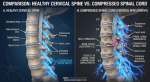
Before discussing surgical candidacy, we must understand the underlying conditions. Cervical radiculopathy occurs when a specific nerve root is pinched. This usually causes sharp, radiating pain down one arm1. Patients often complain of localized tingling and isolated muscle weakness.
Cervical spondylotic myelopathy (CSM) is a much more serious condition. It involves direct physical compression of the main spinal cord2. CSM symptoms progress gradually and can cause permanent paralysis. Patients often present with clumsy hands and frequent stumbling. They may also experience hyperreflexia and dangerous urinary sphincter dysfunction2.
Both conditions can eventually require surgical cervical decompression. However, their distinct clinical presentations dictate entirely different surgical approaches. Accurate diagnosis remains the very first step in assessing candidacy.
Conservative Care: The First Step
Surgery is rarely the first option for cervical nerve compression. Most clinical guidelines recommend exhausting conservative treatments first3. Many patients improve significantly without any surgical intervention.
Conservative care includes structured physical therapy and targeted exercises4. Anti-inflammatory medications and cervical steroid injections are also standard5. Read our comprehensive guide on conservative spine care here. Non-surgical spinal decompression therapy is another viable conservative alternative. Devices like the DRX9000 apply gentle traction to the neck6.
This traction creates negative pressure within the spinal discs. This negative pressure can retract bulging discs away from nerves7. Some studies show significant pain reduction with non-surgical decompression8. However, conservative care is not appropriate for every single patient.
When Conservative Treatment Fails
Patients must be evaluated for surgery when conservative measures fail. The North American Spine Society provides clear clinical surgical guidelines9. Surgery is indicated if severe arm pain remains highly disabling4.
It is also indicated if neurological weakness progressively worsens10. Progressive weakness implies that permanent nerve damage is actively occurring. Surgeons must intervene quickly to prevent irreversible muscle atrophy.
Signs of active spinal cord compression require urgent surgical evaluation4. Myelopathy rarely improves with physical therapy or spinal injections. In fact, delaying surgery for severe myelopathy worsens long-term outcomes. Therefore, profound neurological deficits warrant early and decisive surgical decompression11.
Identifying the Ideal Surgical Candidate
An ideal surgical candidate possesses specific clinical and radiographic traits. The patient must have symptoms directly matching their MRI findings11. If symptoms and imaging do not align, surgery is inappropriate.
Ideal candidates generally have preserved cervical lordosis. Lordosis is the natural, healthy inward curve of the neck. They should also possess strong, dense cervical bone structures. Good bone density allows surgical hardware to anchor securely.
Furthermore, ideal candidates do not have severe axial neck pain. Neck pain alone is rarely cured by decompression surgery11. Arm pain and neurological deficits respond much better to surgery. The surgeon must clearly align patient expectations with realistic outcomes.
Clinical Tools for Assessing Candidacy
Surgeons use objective scoring systems to evaluate disease severity. The modified Japanese Orthopaedic Association (mJOA) scale is universally utilized12. It provides a standardized method to quantify spinal cord dysfunction.
The mJOA scale assesses four specific neurological domains2. These include upper extremity motor function and lower extremity function. It also measures sensory function and urinary sphincter control13. The maximum possible score is eighteen points.
The mJOA Scoring System
| Severity Category | mJOA Score Range | Clinical Implication |
| Normal | 18 | No functional neurological impairment detected. |
| Mild Myelopathy | 15 – 17 | Minor symptoms; conservative care or monitoring may suffice. |
| Moderate Myelopathy | 12 – 14 | Noticeable impairment; surgical decompression is strongly recommended. |
| Severe Myelopathy | 0 – 11 | Profound disability; urgent surgical intervention is absolutely necessary. |
Patients with moderate to severe mJOA scores are primary candidates2. Surgery halts disease progression and often improves these functional scores. Early intervention generally yields much better postoperative mJOA recovery13.
Radiographic Criteria for Candidacy
Surgeons rely heavily on advanced imaging to confirm surgical candidacy. Static Magnetic Resonance Imaging (MRI) is the traditional diagnostic standard. It visualizes soft tissues, herniated discs, and spinal cord changes.
However, static MRI may miss dynamic spinal cord compression. The cervical spine is highly mobile during normal daily activities. Therefore, dynamic flexion-extension MRI is sometimes required14.
Dynamic MRI captures images while the neck is actively bent. It reveals occult compression that vanishes in a neutral position14. Neck extension often exacerbates cervical stenosis and cord compression significantly15. Studies show compression worsens during extension in 85% of cases16.
The Torg-Pavlov Ratio
The Torg-Pavlov ratio helps identify congenital cervical spinal stenosis17. It compares the spinal canal diameter to the vertebral body18. It removes the risk of magnification errors on simple radiographs19.
A ratio below 0.8 indicates significant developmental spinal stenosis18. The normal minimal space available for the spinal cord is 10mm20. Patients with low ratios have less space for their spinal cord.
They are highly susceptible to myelopathy from minor disc bulges17. These patients are often excellent candidates for multi-level decompression procedures.
Sagittal Vertical Axis and Alignment
Global spinal alignment profoundly impacts surgical decision making. Surgeons measure the C2-C7 Sagittal Vertical Axis (SVA) before surgery21. This axis measures how far forward the head rests.
A high SVA means the head is positioned too far forward. This forward posture strains the neck muscles and worsens disability22. A C2-C7 SVA greater than 40 millimeters predicts poor clinical outcomes21.
Decompression alone is not enough for patients with severe misalignment. The surgeon must actively reconstruct and restore the natural cervical lordosis. Correcting alignment relieves muscle tension and improves long-term quality of life.
Physical Examination and Provocative Testing
Surgeons use provocative physical exams to diagnose cervical radiculopathy clinically. The Spurling test is a famous and highly specific diagnostic maneuver23.
The surgeon gently extends and rotates the patient’s neck backward. Applying downward pressure reproduces the patient’s sharp, radiating arm pain24. A positive Spurling test strongly indicates cervical nerve root compression24.
While highly specific, the test has relatively low overall sensitivity25. This means a negative test does not completely rule out radiculopathy. It must be used alongside advanced imaging and detailed neurological assessments.
Other diagnostic signs include the Lhermitte sign and the Hoffmann sign. The Lhermitte sign involves an electric shock sensation upon neck flexion2. The Hoffmann sign indicates upper motor neuron dysfunction and corticospinal involvement2.
Absolute Contraindications to Cervical Decompression
While many benefit from surgery, certain conditions strictly prohibit it. These are known as absolute surgical contraindications. Performing surgery on these patients invites catastrophic hardware failure.
It also risks severe neurological deterioration and life-threatening complications. Surgeons must aggressively screen for these dangerous exclusionary criteria. The most critical contraindications include severe instability, fractures, and osteoporosis. We will explore each of these specific contraindications in detail below.
Contraindication 1: Severe Spinal Instability
Severe spinal instability is a major contraindication for certain procedures. Instability means the vertebrae move abnormally and dangerously during motion. It is often defined as translation greater than three millimeters26.
It can also involve excessive rotational differences between adjacent vertebrae27. If a spine is highly unstable, simple decompression is insufficient. Removing bone to decompress nerves actually worsens the underlying instability.
For example, cervical disc arthroplasty (CDA) is absolutely contraindicated here27. CDA relies on a stable spine to function correctly. Placing an artificial disc in an unstable spine causes mechanical failure. Instead, these patients require rigid spinal fusion to restore stability.
Contraindication 2: Recent Fractures and Trauma
Recent cervical fractures completely alter the surgical decision-making process. Trauma can severely compromise the structural integrity of the spinal column28. A fractured vertebra cannot support artificial discs or simple decompression.
Post-traumatic vertebral body deformity is an absolute contraindication for CDA27. Standalone posterior laminectomies are also contraindicated after severe trauma. Removing the posterior lamina destabilizes an already fractured spinal column.
Patients with recent fractures require highly specialized trauma surgery. This usually involves extensive anterior and posterior instrumented spinal fusion. The primary goal shifts from simple decompression to preventing immediate paralysis.
Contraindication 3: Advanced Osteoporosis
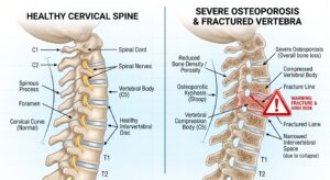
Advanced osteoporosis is perhaps the most dangerous surgical contraindication29. Osteoporosis severely reduces bone mineral density and overall bone strength. Weak bones cannot securely hold titanium screws or fusion cages.
When hardware is placed into osteoporotic bone, it often fails. The most common mechanical complication is known as cage subsidence30. Subsidence occurs when a fusion cage sinks into the weakened vertebra.
This sinking causes a sudden loss of intervertebral disc height. It can recreate nerve compression and alter global spinal alignment30. Severe osteoporosis is therefore an absolute contraindication for artificial discs27.
Assessing Bone Density with Hounsfield Units
Historically, DEXA scans were used to diagnose osteoporosis preoperatively. However, modern spine surgeons now utilize CT Hounsfield Units (HU). Hounsfield units directly measure bone density at the exact surgical site.
Lower HU values correlate strongly with postoperative cage subsidence31. An HU value below 300 indicates poor local bone quality33. Values below 330 are also linked to higher subsidence rates33.
Patients with low HU values require extensive preoperative medical optimization. Surgeons may prescribe osteoporosis medications to improve bone density first32. They may also alter their surgical technique to prevent subsidence.
Hounsfield Unit Predictors for Subsidence
| Hounsfield Unit (HU) Value | Bone Quality Assessment | Risk of Cage Subsidence |
| > 350 HU | Normal / Healthy Bone | Very Low Risk |
| 300 – 350 HU | Osteopenia / Mild Weakness | Moderate Risk |
| < 300 HU | Osteoporosis / Poor Quality | High Risk (Contraindication) |
Contraindication 4: Severe Cervical Kyphosis
Cervical kyphosis is a reversal of the normal neck curve. The neck bends forward unnaturally, creating a structural “hunchback” appearance. Severe kyphosis is a major contraindication for posterior decompression procedures34.
Posterior procedures include laminoplasty and standalone laminectomy. If laminoplasty is performed on a kyphotic spine, it fails35. The spinal cord remains draped over the front of the canal.
Therefore, posterior decompression does not relieve the anterior cord pressure36. Kyphosis greater than 15 degrees requires an anterior surgical approach26. Anterior approaches allow the surgeon to actively correct the spinal deformity.
Differentiating CSM from Mimicking Diseases
Misdiagnosis is a massive risk when evaluating cervical decompression candidates. Cervical spondylotic myelopathy closely mimics other neurological disorders. Surgeons must rule out these diseases before performing any surgery.
Amyotrophic Lateral Sclerosis (ALS)
CSM can closely mimic Amyotrophic Lateral Sclerosis (ALS)37. Both diseases cause profound muscle weakness, atrophy, and stiff limbs. However, CSM is a highly treatable structural spinal problem.
ALS is a fatal, progressive neurodegenerative disease38. Performing spine surgery on an ALS patient provides no benefit. It only subjects a dying patient to unnecessary surgical trauma.
Surgeons must look for specific clinical clues to differentiate them. CSM frequently causes numbness, tingling, and sharp radiating pain37. ALS primarily affects motor nerves, making sensory pain very rare2.
CSM also causes bowel and bladder dysfunction, unlike ALS37. Electromyography (EMG) and careful MRI review remain vital diagnostic tools.
Multifocal Motor Neuropathy (MMN)
Multifocal Motor Neuropathy is another dangerous ALS and CSM look-alike37. It is an autoimmune disorder attacking the body’s motor nerves. MMN causes progressive asymmetric muscle weakness in the hands and arms.
Unlike CSM, MMN rarely causes sensory symptoms or bladder issues37. MMN is highly treatable with intravenous immunoglobulin medications. Proper EMG testing distinguishes MMN from structural cervical cord compression37.
Double Crush Syndrome
Sometimes patients have compression at two entirely different nerve sites. This phenomenon is known as Double Crush Syndrome39. The nerve is pinched in the neck and the wrist simultaneously.
Cervical radiculopathy increases the nerve’s susceptibility to distal carpal tunnel entrapment39. Treating only the neck may not relieve the hand symptoms. Surgeons must carefully evaluate both sites using detailed electrodiagnostic studies40.
Surgical Options for Decompression
When a patient is deemed a candidate, surgeons choose a specific procedure. The choice depends on alignment, pathology location, and bone quality. The most common procedures are ACDF, CDA, and Laminoplasty.
Each procedure has unique candidate criteria and specific biomechanical advantages. Understanding these differences is crucial for optimal surgical decision-making.
Anterior Cervical Discectomy and Fusion (ACDF)
ACDF is considered the traditional gold standard for cervical decompression41. The surgeon approaches the spine from the front of the neck. The damaged disc is entirely removed to decompress the nerves4.
A spacer and bone graft are inserted into the empty space. A titanium plate and screws hold the bones together securely. Eventually, the two vertebrae fuse into one single solid bone.
ACDF is incredibly versatile and effectively treats kyphotic deformities36. It is the absolute best choice for patients with severe spinal instability. However, it permanently eliminates natural motion at the fused segment.
Cervical Disc Arthroplasty (CDA)
CDA is a modern, motion-preserving alternative to traditional spinal fusion. Like ACDF, the surgeon removes the damaged disc from the front. However, instead of fusing the bones, an artificial disc is implanted4.
This artificial implant allows the neck to maintain normal movement11. CDA significantly reduces stress on the adjacent spinal levels11. This reduces the risk of future adjacent segment disease42.
CDA candidates must have isolated one or two-level disc disease. They must not have severe arthritis, instability, or osteoporosis27. Strict patient selection is required for successful artificial disc replacement43.
Posterior Cervical Laminoplasty
Laminoplasty approaches the spinal cord from the back of the neck. The surgeon hinges the lamina bone open like a door44. This instantly expands the spinal canal and relieves cord pressure35.
Small titanium plates keep the bony door permanently propped open. Laminoplasty avoids the mechanical complications associated with spinal fusion. It is ideal for patients with multi-level congenital spinal stenosis36.
However, candidates must have preserved cervical lordosis for it to work35. Patients with severe axial neck pain should absolutely avoid this procedure35.
Laminectomy and Fusion
Laminectomy involves the complete surgical removal of the posterior spinal lamina. Standalone laminectomy is rarely performed due to the risk of kyphosis36. Without the posterior tension band, the neck collapses forward over time.
Therefore, laminectomy is usually combined with posterior instrumented spinal fusion. Screws and rods are placed in the lateral masses for stability. This is highly effective for severe, multi-level posterior compression pathology.
However, posterior fusion carries higher risks of wound healing complications35. It also involves more significant intraoperative blood loss than laminoplasty45.
Endoscopic Cervical Surgery
Endoscopic spine surgery (ESS) is a rapidly emerging, minimally invasive technique. Surgeons use tiny cameras and specialized tools through small incisions. Anterior endoscopic cervical discectomy (AECD) avoids extensive muscle dissection46.
Posterior endoscopic cervical foraminotomy (PECF) is another excellent minimally invasive option. PECF is highly effective for isolated foraminal stenosis and radiculopathy46. It provides comparable arm pain relief to ACDF with faster recovery47.
Endoscopic procedures require highly specialized surgical training and expertise. They are generally not indicated for severe instability or advanced myelopathy46.
Comparing the Surgical Procedures
| Feature | ACDF (Fusion) | CDA (Artificial Disc) | Laminoplasty |
| Approach | Anterior (Front) | Anterior (Front) | Posterior (Back) |
| Motion Preservation | No (Fuses segments) | Yes (Maintains motion) | Partial (Preserves some motion) |
| Best For | Instability, Kyphosis, Trauma | Young patients, single-level disease | Multi-level congenital stenosis |
| Major Contraindications | None specific | Osteoporosis, Instability, Kyphosis | Severe Kyphosis, Severe Neck Pain |
| Key Complication Risk | Adjacent Segment Disease | Implant Subsidence | C5 Nerve Root Palsy |
The Risk of C5 Nerve Root Palsy
Every surgical procedure carries specific neurological and mechanical risks. C5 nerve root palsy is a dreaded complication of cervical decompression. It occurs in up to 30 percent of posterior decompression surgeries48.
Patients wake up with severe weakness in their deltoid and biceps48. They often cannot raise their arms above their shoulders. This palsy usually develops within one to four weeks postoperatively48.
The exact mechanism of C5 palsy remains highly debated among surgeons. It likely involves a rapid backward shift of the spinal cord48. This shift stretches and tethers the delicate C5 nerve root.
Spinal cord ischemia and reperfusion injury also play a role49. Laminoplasty carries a lower risk of C5 palsy than laminectomy50. Circumferential fusion poses the highest overall risk for C5 palsy50.
The Risk of Adjacent Segment Disease
Adjacent segment disease (ASD) is the primary drawback of ACDF surgery. Fusing a spinal segment eliminates all natural motion at that joint. The surrounding spinal discs must absorb extra mechanical stress to compensate11.
This extra stress accelerates the degeneration of these adjacent levels11. Patients may develop new bone spurs and new nerve compression28. Long-term data shows high rates of ASD after traditional fusion51.
Roughly 25 percent of ACDF patients develop ASD within ten years51. Many of these patients eventually require a second revision surgery. This is exactly why CDA is preferred for young, healthy surgical candidates.
10-Year Long-Term Outcomes: ACDF vs CDA
Recent ten-year studies highlight the long-term benefits of motion preservation. CDA demonstrates superior long-term clinical outcomes compared to traditional ACDF52.
The cumulative risk of subsequent surgery is much lower for CDA53. At ten years, ACDF patients undergo significantly more revision surgeries52. ACDF patients also suffer higher rates of long-term mechanical failure52.
CDA maintains high clinical success rates and excellent patient satisfaction53. However, these excellent results depend entirely on strict patient selection criteria. Only perfect candidates achieve these optimal long-term artificial disc outcomes.
Healthcare Economics and Cost-Effectiveness
Healthcare economics heavily influence the choice of surgical procedure today. ACDF and CDA have similar initial surgical costs around $26,00054. However, CDA shows lower global healthcare costs over a two-year period54.
This cost saving is due to fewer readmissions and subsequent procedures. When treating multi-level disease, laminoplasty is highly cost-effective55. Laminoplasty has lower supply and personnel costs compared to posterior fusion55.
ACDF for multi-level disease is associated with the highest overall costs56. Therefore, laminoplasty provides a highly efficient surgical option for severe myelopathy55. Laminectomy with fusion consistently incurs the greatest long-term treatment costs57.
Post-Operative Recovery Expectations
Recovery timelines vary based on the specific surgical approach utilized. Anterior procedures like ACDF and CDA generally feature faster early recoveries. These procedures bypass the thick muscle layers of the posterior neck46.
Patients often return to driving within two to three weeks58. Return to light work is possible within two to four weeks. However, ACDF patients must wait months for the bone to fuse28.
Posterior procedures like laminoplasty require longer muscle healing times. The posterior cervical muscles are crucial for supporting the heavy head. Dissecting these muscles increases postoperative pain and extends the rehabilitation period.
Managing Post-Operative Complications
Despite careful candidate selection, postoperative complications can still occur. Dysphagia, or difficulty swallowing, is common after anterior cervical surgery. This occurs due to retraction of the esophagus during the procedure.
Dysphagia usually resolves spontaneously within a few weeks of surgery. However, persistent dysphagia can significantly impact a patient’s quality of life. Surgeons may employ steroid treatments to reduce local postoperative swelling.
Infection is another risk, particularly with posterior cervical surgical approaches35. The posterior neck has higher rates of surgical site wound complications35. Careful soft tissue management and topical antibiotics help mitigate this risk35.
Pseudarthrosis: The Failure to Fuse
Pseudarthrosis is a specific complication unique to spinal fusion procedures. It means the targeted bones fail to successfully fuse together. This creates a painful, unstable joint known as a “false joint.”
Pseudarthrosis often causes chronic neck pain and hardware loosening over time. It is significantly more common in patients who smoke tobacco. Nicotine severely inhibits bone growth and impairs local blood supply.
It is also highly prevalent in patients with severe unmanaged osteoporosis28. Treating pseudarthrosis is difficult and usually requires a complex revision surgery28. Surgeons may use posterior instrumentation to forcefully stabilize the failed anterior fusion.
Summarizing Surgical Candidacy
Cervical decompression safely restores neurological function in carefully selected patients. The ideal candidate suffers from progressive myelopathy or unrelenting arm pain. Their symptoms must perfectly match advanced MRI and CT imaging findings.
Surgeons must rigorously screen for dangerous contraindications like severe osteoporosis. Unstable spines and kyphotic deformities require complex fusions, not simple decompressions. Misdiagnosing conditions like ALS leads to tragic and unnecessary surgical interventions.
By utilizing scores like the mJOA and analyzing Hounsfield units, surgeons optimize safety. Matching the right procedure to the right patient guarantees long-term success.
Works cited
- NASS Releases Guideline for Diagnosis and Treatment of Cervical, https://www.beckersspine.com/spine/nass-releases-guideline-for-diagnosis-and-treatment-of-cervical-radiculopathy/
- Cervical Myelopathy – StatPearls – NCBI Bookshelf – NIH, https://www.ncbi.nlm.nih.gov/books/NBK482312/
- Posterior decompressive procedures for the cervical spine – PubMed, https://pubmed.ncbi.nlm.nih.gov/12690873/
- Surgery for cervical radiculopathy – Sword Health, https://swordhealth.com/care-explorer/cervical-radiculopathy/surgery
- Cervical Epidural Injections and Diagnostic Spinal Nerve Blocks, https://hoosierpt.com/files/pdf/CervicalEpiduralInjections.pdf
- DRX9000 Research – True Spinal Decompression – Excite Medical, https://excitemedical.com/drx9000-research/
- Effects of Nonsurgical Spinal Decompression Treatment on the, https://www.e-jar.org/journal/view.html?uid=2478&vmd=Full
- Retrospective Chart Review of Nonsurgical Spinal Decompression, https://academic.oup.com/milmed/article/190/Supplement_2/134/8256266
- An evidence-based clinical guideline for the diagnosis … – PubMed, https://pubmed.ncbi.nlm.nih.gov/21168100/
- APA | Cervical radiculopathy – Australian Physiotherapy Association, https://australian.physio/inmotion/cervical-radiculopathy
- Cervical radiculopathy: ‘It’s the decision, not the incision’ that dictates, https://www.mayoclinic.org/medical-professionals/orthopedic-surgery/news/cervical-radiculopathy-its-the-decision-not-the-incision-that-dictates-outcomes/mac-20568261
- The modified Japanese Orthopaedic Association scale – PubMed, https://pubmed.ncbi.nlm.nih.gov/27342612/
- Characterizing mJOA-defined post-surgical recovery patterns in, https://pmc.ncbi.nlm.nih.gov/articles/PMC10772397/
- Use of Flexion-Extension MRI to Reveal Occult Spondylotic, https://pubmed.ncbi.nlm.nih.gov/41229404/
- Radiological and Clinical Significance of Cervical Dynamic, https://pubmed.ncbi.nlm.nih.gov/38955522/
- Use of Flexion-Extension MRI to Reveal Occult Spondylotic, https://www.neurology.org/doi/10.1212/CPJ.0000000000200555
- A New Method for Identifying Predisposition to Spinal Cord Injury, https://pubmed.ncbi.nlm.nih.gov/28807778/
- Torg Ratio for Determining Presence of Spinal Canal Stenosis, https://www.ebmconsult.com/articles/torg-ratio
- The Torg–Pavlov Ratio in Cervical Spondylotic Myelopathy, https://www.researchgate.net/publication/11850563_The_Torg-Pavlov_Ratio_in_Cervical_Spondylotic_Myelopathy_A_Comparative_Study_Between_Patients_With_Cervical_Spondylotic_Myelopathy_and_a_Nonspondylotic_Nonmyelopathic_Population
- A Comparison of Computed Tomography Measures for Diagnosing, https://pmc.ncbi.nlm.nih.gov/articles/PMC4330215/
- A systematic review and meta-analysis of sagittal cervical spine, https://pubmed.ncbi.nlm.nih.gov/41467167/
- The Sagittal Balance of Cervical Spine : Comprehensive Review of, https://pmc.ncbi.nlm.nih.gov/articles/PMC10641420/
- Spurling Test – PubMed, https://pubmed.ncbi.nlm.nih.gov/29630204/
- Spurling Test – StatPearls – NCBI Bookshelf – NIH, https://www.ncbi.nlm.nih.gov/books/NBK493152/
- Diagnostic Performance of Spurling’s Test for the Assessment, https://pubmed.ncbi.nlm.nih.gov/39938056/
- [What are the Criteria for Deciding Posterior Cervical … – PubMed, https://pubmed.ncbi.nlm.nih.gov/34879342/
- Cervical Intervertebral Disk Arthroplasty | Musculoskeletal Key, https://musculoskeletalkey.com/cervical-intervertebral-disk-arthroplasty/
- Cervical adjacent segment disease: Risks and complications, https://caringmedical.com/prolotherapy-news/post-cervical-spine-fusion-surgery-pain-adjacent-segment-disease/
- Osteoporosis and the Management of Spinal Degenerative Disease, https://pmc.ncbi.nlm.nih.gov/articles/PMC5736884/
- Hounsfield Unit Utilization in Cervical Spine for Bone Quality … – PMC, https://pmc.ncbi.nlm.nih.gov/articles/PMC11766049/
- Lower Hounsfield units on CT are associated with cage subsidence, https://pubmed.ncbi.nlm.nih.gov/32503001/
- An analysis of different modalities of bone mineral densitometry, https://pmc.ncbi.nlm.nih.gov/articles/PMC11666519
- The predictive value of Hounsfield units for titanium mesh cage, https://www.frontiersin.org/journals/surgery/articles/10.3389/fsurg.2022.1012364/full
- Surgical treatment of cervical kyphosis – PMC – NIH, https://pmc.ncbi.nlm.nih.gov/articles/PMC3065605/
- Cervical laminoplasty: indication, technique, complications – PMC, https://pmc.ncbi.nlm.nih.gov/articles/PMC7154346/
- Posterior Surgical Techniques for Cervical Spondylotic Myelopathy, https://www.e-neurospine.org/journal/view.php?number=926
- Can Cervical Spondylotic Myelopathy Be Misdiagnosed as ALS?, https://incitefulmed.com/resources/questions/can-cervical-spondylotic-myelopathy-be-misdiagnosed-as-als/
- Amyotrophic lateral sclerosis: frequent complications by cervical, https://pubmed.ncbi.nlm.nih.gov/14648282/
- Carpal tunnel and double crush: comparing outcomes, https://www.handtherapyacademy.com/uncategorized/carpal-tunnel-release-remains-effective-in-patients-with-double-crush-syndrome-and-cervical-radiculopathy/
- The Double Crush Syndrome – PMC – NIH, https://pmc.ncbi.nlm.nih.gov/articles/PMC5823245/
- Cost-utility analysis modeling at 2-year follow-up for cervical disc, https://pmc.ncbi.nlm.nih.gov/articles/PMC4300975/
- a meta-analysis of rates of adjacent-level surgery to 7-year follow-up, https://pmc.ncbi.nlm.nih.gov/articles/PMC7154351/
- The prevalence of indications and contraindications to cervical total, https://www.researchgate.net/publication/5862542_The_prevalence_of_indications_and_contraindications_to_cervical_total_disc_replacement
- Current Diagnosis and Management of Cervical Spondylotic … – Ovid, https://www.ovid.com/journals/glspn/fulltext/10.1177/2192568217699208~current-diagnosis-and-management-of-cervical-spondylotic
- Anterior cervical discectomy and fusion, open-door laminoplasty, or, https://pmc.ncbi.nlm.nih.gov/articles/PMC9879003/
- Current Indications for Spinal Endoscopic Surgery and Potential for, https://pmc.ncbi.nlm.nih.gov/articles/PMC10080449/
- Full-Endoscopic Posterior Cervical Decompression Versus Anterior, https://pmc.ncbi.nlm.nih.gov/articles/PMC13314661/
- C5 Nerve Palsy After Posterior Instrumentation and Decompression, https://pmc.ncbi.nlm.nih.gov/articles/PMC12177111/
- C5 Palsy After Cervical Decompression: A 10-Year Single-Institution, https://pmc.ncbi.nlm.nih.gov/articles/PMC12956150/
- Incidence of C5 palsy in anterior cervical decompression & fusion, https://pmc.ncbi.nlm.nih.gov/articles/PMC12960464/
- Adjacent Segment Pathology After Treatment With Cervical Disc, https://pmc.ncbi.nlm.nih.gov/articles/PMC7343266/
- Increased Cumulative Incidence of 10-Year Reoperation and, https://pubmed.ncbi.nlm.nih.gov/42101246/
- Results From a Prospective, Randomized Clinical Trial at 3 Sites, https://pubmed.ncbi.nlm.nih.gov/37028803/
- Health Care Costs Following Anterior Cervical Discectomy and, https://pubmed.ncbi.nlm.nih.gov/38192187/
- Cervical Laminoplasty Versus Laminectomy and Fusion – PubMed, https://pubmed.ncbi.nlm.nih.gov/39235085/
- Anterior Cervical Discectomy and Fusion Versus Laminoplasty for, https://pubmed.ncbi.nlm.nih.gov/34153482/
- A Systematic Review and Meta-Analysis of Cost and Patient, https://pubmed.ncbi.nlm.nih.gov/41489664/
- E-Posters – PMC, https://pmc.ncbi.nlm.nih.gov/articles/PMC11086047/